Why Your Doctor Recommended an Abdominal Scan: A Patient's Guide
Published: 12 May 2026 | Reviewed by: Gani Hospitals Radiology & Internal Medicine Team
Being advised to undergo an abdominal scan Ramanathapuram may feel worrying, especially when the reason is not fully explained. Most patients visiting our scan center Ramanathapuram have similar concerns and questions. This guide helps you understand everything clearly. An abdominal ultrasound is a safe, painless, and radiation-free test used to examine internal organs in real time. At Gani Hospitals Radiology, our team provides accurate diagnostic imaging Ramnad to help doctors detect conditions early and plan the right treatment. Whether you searched for an ultrasound scan near me or were referred by your doctor, this guide explains what the scan is, why it is needed, and how to prepare for your visit.

An abdominal scan Ramanathapuram at our radiology department uses high-frequency ultrasound to examine the liver, gallbladder, pancreas, spleen, kidneys, bladder, and major blood vessels for abnormalities including stones, cysts, tumours, inflammation, and structural changes. It is the most commonly recommended first-line imaging investigation for abdominal pain, bloating, jaundice, urinary symptoms, and digestive problems. Our scan center Ramanathapuram delivers same-day reports reviewed by a qualified radiologist, with no radiation exposure and no recovery time required after the procedure.
Introduction — What Is an Abdominal Ultrasound Scan?
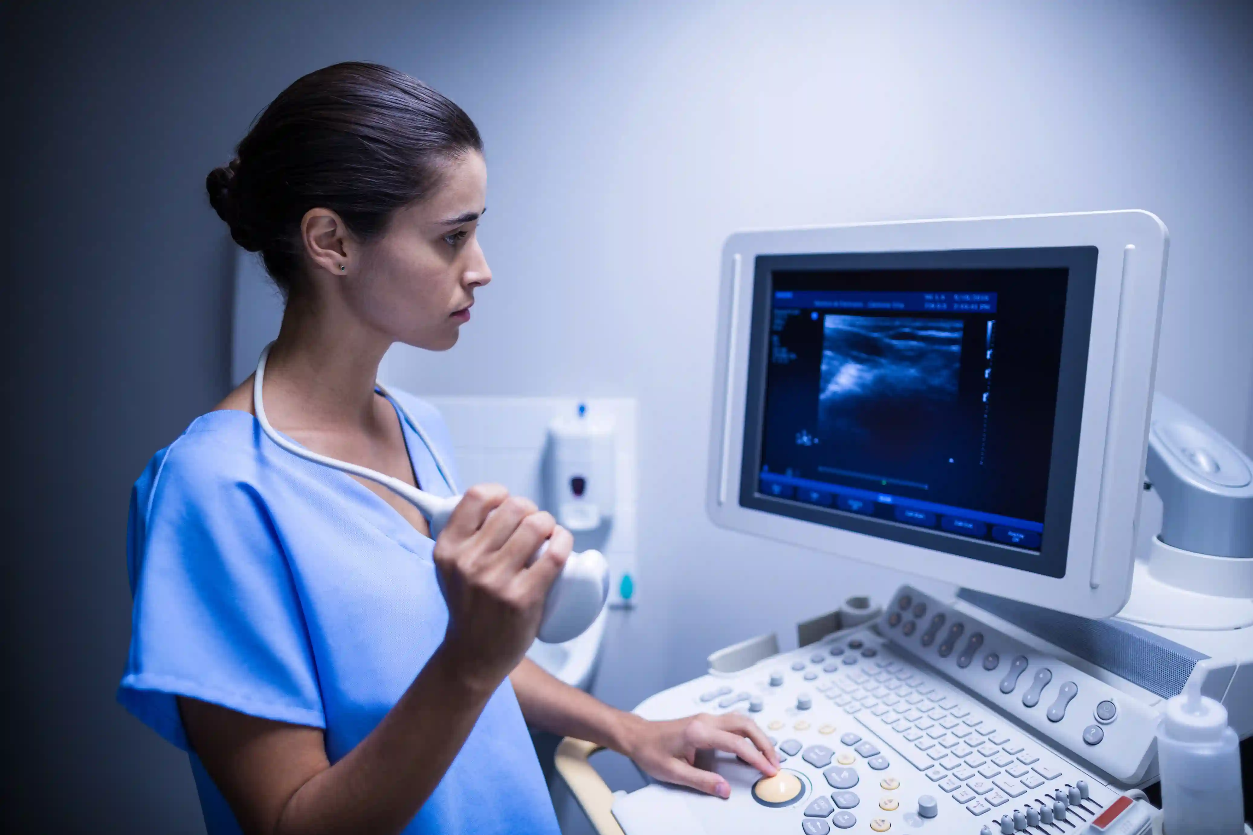
An abdominal ultrasound uses high-frequency sound waves — not X-rays or radiation — to produce real-time images of the organs inside your abdomen. A small handheld device called a transducer is moved gently over the surface of your skin after applying a water-based gel. The sound waves bounce off internal structures and return as echoes that a computer converts into detailed grayscale images on a screen.
When patients search for an ultrasound scan near me, an abdominal scan is among the most frequently performed studies at any diagnostic imaging Ramnad facility. It is fast, painless, safe for all age groups including children and pregnant women, and provides immediate diagnostic information. At our radiology department department, every abdominal scan Ramanathapuram is performed by a qualified sonographer and formally reported by an MD radiologist — ensuring the accuracy and clinical reliability your treating doctor depends upon.
A full abdominal scan typically evaluates eight major organ systems in a single session, making it one of the most information-rich investigations available for the cost and time involved. Our scan center Ramanathapuram is equipped with high-resolution ultrasound machines that capture fine structural detail, Doppler flow assessment, and measurements your physician uses directly in clinical decision-making.
Organs Evaluated During an Abdominal Scan
Liver
Size, texture, echogenicity, fatty change, cysts, lesions, and hepatic vessel flow.
Gallbladder
Gallstones, polyps, wall thickening, bile duct dilatation, and cholecystitis.
Pancreas
Size, echogenicity, ductal dilatation, cysts, and signs of pancreatitis.
Spleen
Size, texture, splenomegaly, infarcts, cysts, and accessory spleens.
Kidneys
Stones, cysts, hydronephrosis, cortical thinning, and renal masses.
Urinary Bladder
Wall thickness, stones, tumours, residual urine volume, and diverticula.
Aorta & Vessels
Abdominal aortic aneurysm screening, vessel calibre, and Doppler flow patterns.
Uterus & Ovaries
In women — uterine fibroids, ovarian cysts, pelvic fluid, and endometrial thickness.
Why Your Doctor Recommended an Abdominal Scan — 10 Common Reasons
Doctors refer patients to a diagnostic imaging Ramnad facility for abdominal scans for a wide range of clinical reasons. Here are the most common situations that lead to a referral to our scan center Ramanathapuram:
Abdominal Pain
Pain in the upper right abdomen often points to gallstones or liver pathology. Left-side pain may indicate the spleen, kidney, or colon. Central pain around the navel can suggest the pancreas, aorta, or mesenteric nodes. An abdominal ultrasound Ramnad study identifies the source quickly and non-invasively.
Jaundice
Yellow discolouration of the skin or eyes is caused by elevated bilirubin. An abdominal scan examines the liver, bile ducts, and gallbladder to determine whether the jaundice is due to gallstones blocking the bile duct, hepatitis, cirrhosis, or a pancreatic mass compressing the common bile duct.
Abnormal Liver Function Tests
If your blood tests show elevated SGOT, SGPT, or bilirubin, your doctor will request a liver scan Ramanathapuram to visualise the liver directly. Fatty liver, hepatitis, cirrhosis, and liver lesions are all identifiable on ultrasound even when symptoms are absent.
Burning or Frequent Urination
Urinary symptoms including burning, frequency, difficulty voiding, or blood in the urine often prompt a kidney ultrasound Ramnad study. Kidney stones, bladder stones, urinary tract infections with hydronephrosis, and bladder tumours are all detectable through the scan.
Bloating and Indigestion
Persistent abdominal bloating, fullness after meals, and chronic indigestion that does not respond to standard treatment may signal gallbladder disease, fatty liver, pancreatic pathology, or free fluid in the abdomen — all assessed through an abdominal scan Ramanathapuram at our our radiology department department.
Elevated Creatinine or Kidney Function Abnormality
Rising creatinine and urea levels in blood tests alert doctors to declining kidney function. A kidney ultrasound Ramnad assesses kidney size, cortical thickness, obstruction to urine flow, and cystic changes that explain the functional decline and guide treatment decisions.
Pelvic or Lower Abdominal Pain in Women
Women with lower abdominal pain, irregular periods, or suspected ovarian cysts are referred for a pelvic and abdominal ultrasound. At our sonography center near me in Ramanathapuram, our radiologists evaluate the uterus, ovaries, and fallopian tube regions for fibroids, cysts, endometriosis indicators, and ectopic pregnancy.
Diabetes Monitoring
Diabetic patients commonly develop fatty liver, kidney disease, and gallbladder abnormalities as long-term complications. Annual abdominal ultrasound Ramnad studies are recommended for diabetic patients to monitor these complications before they cause symptoms or functional impairment.
Suspected Mass or Lump
When a doctor palpates an abdominal mass during physical examination, or a patient reports feeling a lump, an abdominal scan Ramanathapuram is the first-line investigation. It differentiates between solid and fluid-filled structures, benign and suspicious lesions, and guides whether further imaging such as CT or MRI is required.
Hypertension Evaluation
Uncontrolled or secondary hypertension can originate from the kidneys — a condition called renovascular hypertension. Our our radiology department team performs renal Doppler studies as part of the hypertension workup, assessing blood flow to both kidneys and identifying renal artery stenosis that may be driving the elevated blood pressure.
Types of Abdominal Scan Available at our radiology department
Our our radiology department department offers a full range of abdominal imaging modalities at our scan center Ramanathapuram. Your doctor will specify which type is appropriate based on your clinical presentation:
Standard Abdominal Ultrasound
The most common study — evaluates all major abdominal organs in a single session. The first-line investigation for most abdominal complaints referred to our diagnostic imaging Ramnad centre. Safe, painless, no radiation.
Pelvic Ultrasound
Specifically examines the uterus, ovaries, and pelvic structures in women, and the prostate and bladder in men. Often performed as a combined abdominal and pelvic study at our sonography center near me.
Doppler Ultrasound
Adds blood flow assessment to standard imaging using colour and spectral Doppler technology. Essential for evaluating renal artery stenosis, portal hypertension, hepatic vein thrombosis, and vascular masses. Available at our our radiology department department.
Kidney and Urinary Tract Ultrasound (KUB)
Focused study of both kidneys, ureters, and the urinary bladder. The standard investigation for kidney ultrasound Ramnad referrals covering stone disease, hydronephrosis, renal masses, and bladder pathology.
Liver and Hepatobiliary Scan
Detailed study of the liver, gallbladder, and bile ducts. Used when a liver scan Ramanathapuram referral is made specifically for jaundice evaluation, abnormal LFTs, suspected fatty liver, cirrhosis staging, or hepatic mass characterisation.
High-Resolution Abdominal Ultrasound
Uses higher frequency transducers for more detailed evaluation of superficial abdominal structures including the appendix, bowel wall, mesenteric lymph nodes, and hernia sacs. Requested when standard abdominal ultrasound Ramnad requires supplementary detail.
Conditions Commonly Detected Through Abdominal Ultrasound in Ramanathapuram
The following are the most frequently identified findings at our scan center Ramanathapuram during abdominal ultrasound studies. Early detection through routine or symptomatic diagnostic imaging Ramnad allows for timely treatment and prevents serious complications.
| Organ | Condition Detected | Significance |
|---|---|---|
| Liver | Fatty liver (Grade I, II, III), hepatitis, cirrhosis, liver cysts, hepatic haemangioma, focal lesions | Early cirrhosis and fatty liver are reversible with intervention. Focal lesions require follow-up imaging or biopsy. |
| Gallbladder | Gallstones, biliary sludge, gallbladder polyps, cholecystitis, contracted gallbladder, bile duct stones | Untreated gallstones can cause acute cholecystitis, pancreatitis, and bile duct obstruction requiring emergency surgery. |
| Pancreas | Pancreatitis (acute and chronic), pancreatic cysts, duct dilatation, focal pancreatic masses | Pancreatic masses have a high malignancy risk. Early detection significantly improves surgical outcomes. |
| Spleen | Splenomegaly (enlarged spleen), splenic cysts, splenic infarcts, accessory spleen | Splenomegaly indicates systemic illness including liver disease, haematological conditions, or infection. |
| Kidneys | Renal stones, hydronephrosis, renal cysts, cortical thinning, renal masses, polycystic kidney disease | Obstruction and stones cause progressive kidney damage if untreated. Early kidney ultrasound Ramnad enables timely urology referral. |
| Bladder | Bladder stones, bladder wall thickening, bladder tumours, post-void residual volume, diverticula | Bladder tumours detected early have a much better prognosis than those found at symptomatic stages. |
| Aorta | Abdominal aortic aneurysm (AAA), atherosclerotic plaques, aortic dilatation | AAA can rupture fatally if undetected. Ultrasound screening identifies candidates for surgical repair before rupture. |
| Uterus & Ovaries | Uterine fibroids, ovarian cysts, polycystic ovaries, endometrial thickening, pelvic free fluid | PCOS, fibroids, and endometrial changes affect fertility and long-term hormonal health — all identifiable through our radiology services Ramanathapuram. |
| Lymph Nodes | Mesenteric lymphadenopathy, retroperitoneal lymphadenopathy | Enlarged lymph nodes may indicate infection, inflammatory bowel disease, or lymphoma requiring further evaluation. |
| Free Fluid | Ascites, pelvic free fluid, loculated collections | Ascites indicates advanced liver disease, cardiac failure, malignancy, or peritoneal infection — each requiring distinct treatment pathways. |
Why Choose our radiology department for Your Abdominal Scan in Ramanathapuram
MD Radiologist Reports
Every abdominal scan Ramanathapuram at our centre is formally reported by a qualified MD radiologist — not just a technician-generated image.
High-Resolution Equipment
Our our radiology department machines deliver sharp, detailed images across all frequencies — improving diagnostic accuracy for small lesions and subtle pathology.
Same-Day Reports
Reports from our our imaging centre are issued the same day, shared digitally, and formatted for direct use in specialist referrals and treatment planning.
Colour Doppler Available
Advanced Doppler assessment for vascular evaluation is available at our diagnostic imaging Ramnad centre — essential for renal, hepatic, and portal vein studies.
Hospital-Integrated Reporting
Findings from your abdominal ultrasound Ramnad study feed directly into specialist consultations, inpatient care, and surgery planning within the same hospital.
Accessible Location
Centrally located in Ramanathapuram, our sonography center near me is easily reachable from Rameswaram, Paramakudi, Mandapam, Keelakarai and all surrounding areas.
Patient Privacy
All scans at our radiology services Ramanathapuram centre are conducted in private, enclosed examination rooms with same-gender sonographer available on request.
Transparent Pricing
Clear, upfront scan fees with no hidden charges. Insurance and government health scheme patients are supported at our our radiology department counter.
Walk-In and Appointment
Both walk-in and pre-booked scan appointments accepted at our our imaging centre. Urgent scans for inpatients and emergency referrals prioritised.
How to Prepare for Your Abdominal Ultrasound Scan
For a Full Abdominal Scan
- Fast for 6 to 8 hours before the scan: Food and drink cause the gallbladder to contract and bowel gas to increase, both of which significantly reduce image quality. Plain water is permitted during the fasting period.
- Avoid milk, tea, coffee, and fruit juices: These stimulate gallbladder contraction and impair visualisation of gallbladder stones and the biliary system.
- Avoid gas-producing foods the previous evening: Beans, lentils, carbonated drinks, and raw vegetables increase intestinal gas that can obscure pancreatic and retroperitoneal structures.
- Wear comfortable, loose clothing: Garments that expose the abdomen easily allow the sonographer to position the probe without restriction during your abdominal ultrasound Ramnad study.
For a Pelvic or Lower Abdominal Scan (Including KUB)
- Arrive with a full bladder: Drink 4 to 6 glasses of water one hour before the scan and do not urinate until after the pelvic component is complete. A full bladder acts as an acoustic window that allows clear visualisation of the uterus, ovaries, prostate, and bladder wall.
- If both abdominal and pelvic scans are requested: Our our radiology department team will first perform the pelvic scan with a full bladder, then ask you to empty your bladder for the abdominal component where an empty bladder is preferred.
General Instructions for All Scan Patients
- Bring your doctor's referral letter or scan requisition slip to our our imaging centre reception.
- Carry any previous scan reports or imaging from other facilities — comparison with prior studies significantly enhances diagnostic value.
- Inform the sonographer of any previous abdominal surgery, known medical conditions, or medicines you are currently taking before the scan begins.
- No special preparation is needed for emergency or urgent scans — our radiology services Ramanathapuram team will adapt the protocol based on your clinical situation.
What Happens During Your Abdominal Scan — Step by Step
Many patients feel anxious simply because they do not know what to expect. Here is exactly what happens during an abdominal ultrasound in Ramanathapuram at our our radiology department centre:
Registration
Submit your referral at reception. Our team confirms the scan type and collects your details.
Positioning
You lie on a padded examination couch. Clothing is moved to expose the abdomen. No gown change required in most cases.
Gel Application
A clear, water-based gel is applied to your skin. The gel is warm and washes off easily after the scan.
Scanning
The sonographer moves the transducer over your abdomen, capturing images of each organ systematically. Takes 15 to 30 minutes.
Report Issue
Images reviewed by our MD radiologist. A written report is issued the same day and shared digitally with the patient and referring doctor.
The entire visit to our our imaging centre — from registration to receiving your report — typically takes 45 minutes to one hour for a standard full abdominal study. No recovery time is needed and you may eat and resume normal activities immediately after your diagnostic imaging Ramnad appointment.
Understanding Your Abdominal Scan Report
A formal radiology report from our radiology department follows a structured format. Understanding the terminology helps you engage more meaningfully with your treating doctor when they explain the findings:
- Echogenicity: How bright or dark a structure appears on ultrasound relative to surrounding tissue. Increased echogenicity of the liver suggests fatty infiltration. Decreased echogenicity may indicate hepatitis or oedema.
- Hyperechoic / Hypoechoic / Anechoic: Hyperechoic structures appear bright (such as gallstones). Hypoechoic structures appear dark relative to surroundings. Anechoic means no internal echoes — typically fluid-filled structures like simple cysts.
- Acoustic shadowing: A dark shadow behind a bright structure, characteristic of gallstones and kidney stones where the stone blocks sound from passing through.
- Hydronephrosis: Dilatation of the renal collecting system due to obstruction — graded mild, moderate, or severe. A key finding in kidney ultrasound Ramnad studies for stone disease.
- Splenomegaly: Enlargement of the spleen beyond normal dimensions. Reported with measurement and associated findings that suggest the likely cause.
- Free fluid: Fluid found outside normal anatomical compartments — in the peritoneal cavity (ascites) or pelvis (pelvic free fluid). Always reported with volume estimate and distribution.
- Impression / Conclusion: The final paragraph of the report summarises the significant findings and suggests whether further imaging or clinical correlation is recommended. This section is what your doctor most relies upon.
When Should You Get an Abdominal Scan Without a Doctor's Referral?
While most abdominal ultrasound Ramnad studies are performed following a doctor's referral, there are circumstances where adults are encouraged to proactively request an abdominal scan at our sonography center near me as part of preventive health screening:
- Adults above 40 with a family history of gallstones, kidney stones, liver disease, or abdominal aortic aneurysm
- Diabetic patients who have not had an abdominal scan in the past year — fatty liver and early nephropathy detection are important complications to monitor
- Heavy alcohol consumers — annual liver scan Ramanathapuram monitoring for fatty liver, fibrosis, and early cirrhosis
- Men above 55 who smoke — abdominal aortic aneurysm screening is specifically recommended by international guidelines for this group
- Women above 35 with irregular periods, pelvic pain, or suspected ovarian pathology who have not yet consulted a gynaecologist
- Patients on long-term medications including methotrexate, amiodarone, or corticosteroids — annual liver ultrasound monitors for drug-induced hepatic damage
Self-referred scans at our radiology services Ramanathapuram centre are accepted. Our duty radiologist can advise on follow-up or specialist referral if significant findings are identified during a self-referred radiology imaging in Ramnad study.
Trusted Resources for Ultrasound and Abdominal Imaging Information
For reliable, evidence-based information on abdominal ultrasound, radiology, and diagnostic imaging, refer to these globally trusted health authorities:
- World Health Organization (WHO) — Medical Imaging and Diagnostics
- RadiologyInfo.org — Abdominal Ultrasound Patient Guide (American College of Radiology)
- MedlinePlus — Abdominal Ultrasound Explained
- CDC — Medical Imaging and Radiation Safety
These resources complement the expert clinical guidance provided by our MD radiologists at our radiology department — the most trusted our imaging centre for radiology imaging in Ramnad across the entire district.
Conclusion — Expert Abdominal Imaging When You Need It Most
An abdominal scan is not something to fear — it is one of the most valuable tools modern medicine has for looking inside the body quickly, safely, and accurately. When your doctor recommends an abdominal ultrasound in Ramanathapuram, they are requesting information that will directly guide your diagnosis and treatment. Choosing the right facility matters enormously. A high-resolution machine operated by an experienced sonographer and reported by a qualified radiologist makes the difference between a definitive diagnosis and an inconclusive study that delays your care.
At our radiology department department — the most advanced our imaging centre in the Ramnad district — every radiology imaging in Ramnad study is performed with clinical precision, reported by an MD radiologist the same day, and integrated into the full spectrum of specialist medical care available under one roof. Whether you are a walk-in patient searching for an ultrasound scan near me or a referred patient from your family physician, our radiology services Ramanathapuram team is ready to provide the clarity your doctor needs and the answers you deserve.
Also explore: Pregnancy Scan and Maternity Services at Gani Hospitals | Full Body Health Checkup Packages | Home Blood Test Collection Service | Book a Scan Appointment at Gani Hospitals
Book Your Abdominal Scan Today
Same-day reports. MD radiologist review. Advanced imaging technology.
our radiology department — the most trusted
our imaging centre for radiology imaging in Ramnad.
Frequently Asked Questions
Where can I get an abdominal scan in Ramanathapuram?
The most trusted ultrasound scan near me in Ramanathapuram is at our radiology department department. Our our imaging centre offers comprehensive abdominal ultrasound in Ramanathapuram studies including full abdominal, pelvic, KUB, Doppler, and liver-specific scans with same-day MD radiologist reports.
Why did my doctor recommend an abdominal scan?
Doctors request an abdominal ultrasound Ramnad study for many reasons including abdominal pain, jaundice, abnormal blood tests, urinary symptoms, suspected gallstones or kidney stones, fatty liver monitoring, pelvic pain in women, and hypertension workup. It is the safest first-line radiology imaging in Ramnad investigation for most abdominal complaints.
Is an abdominal ultrasound scan safe?
Yes. Abdominal ultrasound uses sound waves, not radiation, and is completely safe for all patients including children, pregnant women, and elderly individuals. Every abdominal ultrasound in Ramanathapuram at our our radiology department centre is performed following international safety protocols with no known side effects.
Do I need to fast before an abdominal scan?
Yes, for a full abdominal scan a 6 to 8-hour fast is required before your visit to our our imaging centre. This ensures the gallbladder is distended and bowel gas is minimised for the best image quality. For a pelvic or KUB scan, a full bladder is required instead of fasting. Our our radiology department team provides specific instructions when you book your appointment.
How long does an abdominal scan take at Gani Hospitals?
A standard full abdominal ultrasound in Ramanathapuram at our radiology services Ramanathapuram centre takes 15 to 30 minutes for the scanning itself. Including registration and report issuance, most patients complete their entire visit within one hour. Reports are shared digitally the same day.
Can I book an abdominal scan without a doctor's referral?
Yes. Self-referred patients are accepted at our sonography center near me in Ramanathapuram. Our duty radiologist at our radiology department can assess your scan findings and advise on whether specialist follow-up is required. Walk-in and pre-booked appointments are both available at our our imaging centre.